

Spine Surgery
At Brain and Spine Surgery, we also handle different forms of spine treatments, including spine cord tumor removal, minimally invasive spine surgery, and spinal canal stenosis. Our spine doctor in Delhi has years of experience in treating patients suffering from different problems in their spine, which is why we are able to handle multifarious cases. Our professional team not only leverages the modern surgical practices but also implements latest technological innovations like robotic motor arms and GPS-controlled surgical invasions to increase the success rate of every surgery.
We have become the best spine surgery hospital in the capital region owing to our sheer dedication towards treating every patient that walks through the door. Our doctors aim towards minimally invasive surgeries mostly to increase the recovery rate and allow the patients commence their normal lifestyle at the earliest. Apart from this, we pay close attention to the post-surgery period so that we can treat any complication with urgency and reduce the chances of health worsening.
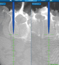
O Arm Guided Spinal Fixation
The O-Arm navigation system is used to guide surgeons for fixing pedicle screws during spine surgeries. It offers real-time 3D images of the body parts and the positioning of the nerves originating from the spinal cord, thereby allowing professionals to accurately fixate the screw without any potential perforation or improper location assessment. With this advanced imaging technology, the success rate of spinal fixation can be greatly increased while reducing any post-surgery complication.
Spine Cord Tumor
As the name suggests, these tumors originate in the spinal cords when cells start dividing at an uncontrollable rate. Primary tumors originate in the tissues of the spinal cord whereas secondary tumors originate in other organs and later travel to the spinal cord through metastasis. Apart from this classification, these can also be grouped based on their locations, namely intramedullary and intradural-extramedullary tumors. The most common symptoms of spinal cord tumors include motor and sensory problems, vertebral collapse, pain, and difficulty in walking.
Intradural Tumor
Intradural tumors originate in the spinal cord or the protective sheath covering the same, also known as the durameter. These are divided into two categories, namely extramedullary and intramedullary depending on the location. Intramedullary tumors originate in the thoracic, cervical, or lumbar regions and are mostly malignant, like gliomas. On the contrary, extramedullary tumors are mostly benign, like meningiomas and nerve sheath tumors. Usually, surgery is preferred for complete removal of the neoplasm or reduction of the size as per the condition of the tumor and its growth rate.
Metastatic Spine Tumor
When the tumor originates in any other part of the body but travels to the spine, it can be termed as the metastatic spine tumor and is considered as the most common form of neoplasms. Spinal metastases can be usually found in the lumbar and thoracic region of the vertebral column. The major symptoms of this condition include weakness in the upper and lower limbs, loss of mobility and difficulty in walking, excruciating pain, and sensory impairment. Also, spinal metastases often lead to nerve root compression, causing myelopathy or radiculopathy.
Minimal Invasive Spine Surgery
In minimally invasive spine surgery, surgeons won’t have to enter the inside of the spinal cord through a large incision. For instance, in endoscopic spine surgery, a thin endoscope is used to gain access into the spine and observe the cells and their current conditions for accurate planning of the surgery and assessment of the progress. Other types of minimally invasive spine surgery include lumbar posterior decompression and anterior lumbar interbody fusion.
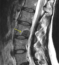
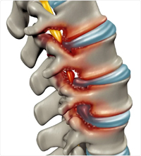
Cervical Disc Prolapse
Cervical disc prolapse is often termed as herniated or slipped disc and occurs when the gel-like cushion padding between two vertebrae pushes through a small tear or perforation in the outer wall. Usually, the herniated sic compresses the nerve roots as it starts protruding out, thereby causing immense pain in the neck and arm. The major symptoms associated with cervical disc prolapse include pain in the shoulder blade, radiating pain in the arms, weakness and tingling sensation in the upper limbs, and loss of coordination and balance.
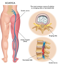
Lumbar Disc Prolapse
Similar to cervical disc prolapse, lumbar disc prolapse occurs in the lumbar vertebrae when the gel-like padding extrudes out of the exterior disc wall through a minute tear. Several events can lead to herniated discs in the lumbar region, including congenital disorder, degeneration of the tissues, sudden trauma while lifting heavy objects, falling from a height, obesity, and poor spinal postures. Conservative treatment options are usually preferred for lumbar disc prolapsed, like administration of pain killers, relaxation, appropriate body positioning, and physiotherapy exercises.
Spinal Canal Stenosis
In spinal stenosis, the spinal canal narrows significantly, causing the nerve roots to compress under immense pressure. It causes excessive pain and numbness in the affected regions, accompanied by tingling sensation, weakness in the limbs, and so on. Spinal stenosis often affects the lumbar and cervical regions of the vertebral column, which is why the upper and lower extremities are impacted first. Treatments often include physical therapies, administration of anti-inflammatory medicines, ligament removal from lumbar spine, and steroid shots.
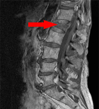
Spinal Fracture
As the name implies, spinal fracture is a condition described when one of the thirty-three bones in the entire vertebral column breaks or suffers from a crack due to trauma. Vertebral compression fractures are categorized to be less painful when compared to other forms, like fracture dislocations and burst fractures. If the fracture is severe, surgery needs to be performed for bone alignment and treatment of the impacted tissues. Also, several patients suffering from minimal spine fracture need to wear braces for months for correct alignment of the bones.
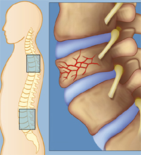
Vertebral Collapse
Also known as vertebral compression fraction (VCF), it is characterized by sudden collapse of the bony structure of the vertebrae in the thoracic region. Symptoms often include sudden onset of pain in the back region, increased pain intensity during mobility, eventual loss in height, limited movement of the spine, and disability or deformity. Osteoporosis is one of the major cases of VCFs since the bony structure starts losing the thickness and integrity and soon collapses.
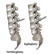
Kyphoplasty / Vertebroplasty
To treat vertebral compression fracture or VCF, doctors can often use two of the most common methods, namely kyphoplasty and vertebroplasty. Usually, in vertebroplasty, doctors use imaging guidance to inject a cement mixture directly into the compressed bone while in kyphoplasty, a balloon is used to create space between the collapsed vertebrae followed by injection of the cement mixture. This cement mixture is usually made from an ingredient termed as polymethylmethacrylate, which hardens as soon as it enters the body.